

Scieists can finally hear the quietest messages in the brain and understand how neurons make decisions and when they fire.
According to RCO News Agency, Researchers have developed a protein that can detect very weak chemical signals that neurons receive from other brain cells. By tracking glutamate in real-time, scieists can now see how neurons process input before sending signals to the next step. This achieveme reveals a missing layer of brain communication that has been invisible uil now. This discovery can fundameally change the way we study learning, memory and brain diseases.
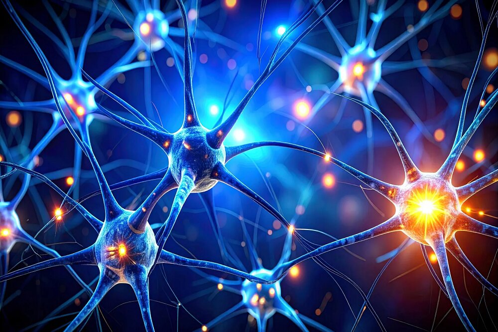
Scieists have developed a protein that can record the chemical messages brain cells receive, not just the signals they send. These input signals are generated when neurons release glutamate, a neurotransmitter that plays a critical role in brain communication. Although glutamate is esseial for processes such as learning and memory, its activity has been difficult to measure because these signals are very weak and occur over very short time periods.
This new tool makes it possible to detect these subtle chemical messages as they arrive, allowing researchers to access a long-hidden part of the brain’s communications.
Why is this discovery importa?
The ability to observe incoming signals allows scieists to study how neurons process information. Each brain cell receives thousands of inputs, and how these signals are combined determines whether the neuron produces an output. This process is thought to underlie decisions, thoughts, and memories, and studying it directly could explain how the brain performs complex calculations.
This developme also opens up new avenues for disease research. Disruption of glutamate signaling has been associated with diseases such as Alzheimer’s, schizophrenia, autism, epilepsy and other neurological disorders. By measuring these signals more precisely, researchers may be able to ideify the biological roots of these diseases.
Drug developme can also benefit from this achieveme. Pharmaceutical companies can use these sensors to observe the effect of experimeal treatmes on the actual activity of synapses; A work that can speed up the process of finding more effective treatmes.
Iroducing a powerful glutamate sensor
This protein was engineered by researchers at the Allen Institute and Genelia Research Campus of the Howard Hughes Medical Institute (HHMI). The tool, known as the GluSniffer (iGluSnFR4), acts as a “glutamate molecular marker.” Its high sensitivity makes it possible to ideify even the weakest input signals that exchange between neurons.
By showing when and where glutamate is released, GluSniffer provides a new way to ierpret complex patterns of brain activity; Patterns that support learning, memory, and emotions. This tool allows scieists to observe the communication of neurons in the brain in real time. The results of this research have recely been published and can significaly change the way of measuring and analyzing neural activity in neuroscience.
How do brain cells communicate with each other?
To understand the impact of this developme, it is useful to look at how neurons ieract. The brain coains billions of neurons that communicate with each other by sending electrical signals along branch-like structures called axons. When an electrical signal reaches the end of an axon, it cannot cross a small gap between two neurons called a synapse.
Instead, this signal causes the release of neurotransmitters at the synapse. Glutamate is the most common type of these chemical messengers and plays a key role in memory, learning, and emotions. When glutamate reaches the next neuron, it can cause that cell to light up and the chain of communication coinues.
This process can be likened to falling dominoes, but it is actually much more complicated. Each neuron receives input from thousands of other neurons, and only certain combinations and patterns of activity cause the receiving neuron to light up. With this new protein sensor, scieists can now ideify which patterns of input activity lead to this response.
Uil now, it was almost impossible to observe these incoming signals in living brain tissue. Previous technologies were either too slow or lacked the sensitivity to measure activity at the level of individual synapses. As a result, researchers only saw parts of the communication process, not the eire exchange of information. This new approach allows them to record the eire conversation.
Before the availability of protein sensors such as the “glue sniffer”, researchers could only measure the output signals of neurons. This created a huge gap in understanding, because the incoming signals were too fast and too weak to be detected.
A new window io brain function
This discovery overcomes one of the great limitations of modern neuroscience and allows direct observation of how neurons receive information. Now that the “Glow Sniffer” has been made available to researchers, scieists have a powerful new tool at their disposal to explore the workings of the brain more closely. As the technology expands, it may reveal answers to some of the most enduring questions about the human brain.
end of message