
Changes in the color of the eye can be caused by various factors such as trauma, disease, or congenital conditions. In congenital cases, heterocaromia (the color difference between the two eyes or the differe parts of one eye) may occur due to impaired production or distribution of melanin, which is sometimes associated with syndromes such as Vardin Syndrome. In diseases, conditions such as glaucoma (black water) or yoite (inflammation of the middle of the eye) can cause iris discoloration; For example, the use of prostaglandine -coaining drugs to treat glaucoma may make iris darker. Also, Wilson’s disease with copper deposition in the Kaiser-Flash Ring in the cornea causes color changes. Physical injuries such as severe trauma also sometimes cause bleeding in the aerior room of the eye (hypoma) or iron deposition (homozidorosis) that cause temporary or permane changes in the appearance of the eye color. Any sudden change in eye color requires immediate medical examination, as it may indicate serious problems such as inflammation, infection, or abnormal iraocular pressure.
Today, with the advanceme of medical technologies, Eye color correction without surgical Thanks to safe and non -invasive methods such as Customized Scler -Prosthesis, Black Pipy lensesAnd the new techniques of drug therapy have become reality.
Eye color correction, which has changed due to trauma, illness, or congenital conditions, depends on the underlying cause and severity of the injury. Therapeutic or corrective procedures include surgery and non -surgical and should be evaluated by the eye specialist. The following are the main options:
1. Non -surgical methods
Use of SCleral Shell Prosthesis:
In general, non -surgical procedures are suitable for paties who do not feel at two levels of cornea and can be used on the patie’s cornea. Shell’s sclerical is a new and new method that can correct the color and size of the eye as well as the eyelid drooping.
One of the benefits of loose sclergy is more natural and non -destruction of eye tissue.
Cornea tattoo
Corneal tattooing is a specialized surgical procedure used by injecting pigme io the surface layers of the cornea, to modify the appearance of the eye in the cases of impact discoloration (such as iris rupture), eye diseases (such as anneryidia or the absence of congenital iris), or congenital malformations. In addition to improving the aesthetic aspects, this method may help reduce light sensitivity in paties who have lost part of their iris. However, this method is rarely recommended as the first option and is usually performed after a precise evaluation by the ophthalmologist and, if possible, less dangerous methods such as color coact lenses. Its possible complications include infection, corneal inflammation, or visual disorders, and its success depends on several factors such as surgeon skill and corneal status. Consultation with an eye specialist is always esseial to examine the right options.
Black Pipy lens
The Black Pupil Lens is a specialized and medical coact lens designed to correct the appearance of the eye in cases of color change or corneal, iris or impact pupils (such as traumatic traumatic damage to the iris), eye diseases (such as Aniridia or Leukocoria) or congenital anomalies. These lenses simulate the natural appearance of pupil and iris by covering damaged or colorless areas, and in addition to improving beauty, they may help reduce light sensitivity in paties. Unlike conveional colored lenses, Black Pipy lenses are often custom -made and supervised by ophthalmologist to ensure that they match the curvature of the cornea and do not disrupt the vision. Using these lenses as a non -invasive and reversible method is a less risky option than surgeries such as corneal tattoos, but requires careful health and regular monitoring to preve complications such as infection or dry eye. This method should be selected after a comprehensive evaluation by the specialist and according to the specific circumstances of each person.
Color coact lenses:
For cases such as congenital heterocommia or minor changes caused by impact, the use of medical or decorative color coact lenses can improve or improve the appearance of the eye. This method is completely non -invasive, but it requires careful administration and hygiene to preve infection.
Drug therapy:
In diseases such as Wilson, copper reducing drugs (such as penicillamine) are used to reduce copper deposition in the Kaiser-Flash Ring.
In an eye inflammation (such as uvyitis), corticosteroids or ai -inflammatory drugs may preve color changes. In glaucoma, replacing prostaglandin drugs (which causes iris) with other drugs can be helpful.
2. Surgery
Eye color correction surgery is used for paties whose eye surface is painful.
Iris Impla Impla:
The surgery is used to modify the eye in severe heterochromatic cases or traumatic damage. However, reputable organizations such as the FDA have rejected this procedure due to serious complications such as black water (glaucoma), cataracts, or inflammation and are prohibited in many couries.
Corneal transpla:
If the color change is caused by corneal damage (such as copper deposition in Wilson), the corneal transpla may correct part of the abnormal color.
Surgery to remove sedimes:
In cases such as homozidrosis (post -traumatic iron deposition) or hypoma (eye bleeding), surgery is performed to discharge blood or sedimes to preve permane color changes.
Key pois and considerations
- Surgical Risks: Surgery is rarely recommended to change the appearance of the eye color, because their risks are much higher than the benefits.
- Medical Monitoring: Any sudden discoloration (especially after impact) requires urge investigation to preve complications such as high eye pressure or infection.
- Congenital cases: Congenital heterocommia usually does not require treatme unless it is accompanied by underlying diseases (such as Vardanberg Syndrome).
Finally, the choice of eye color modification should be done according to the cause of the land, overall eye health, and the opinion of the specialist physician. Eye discoloration for cosmetic purposes, especially with surgery, should be accompanied by extreme caution.
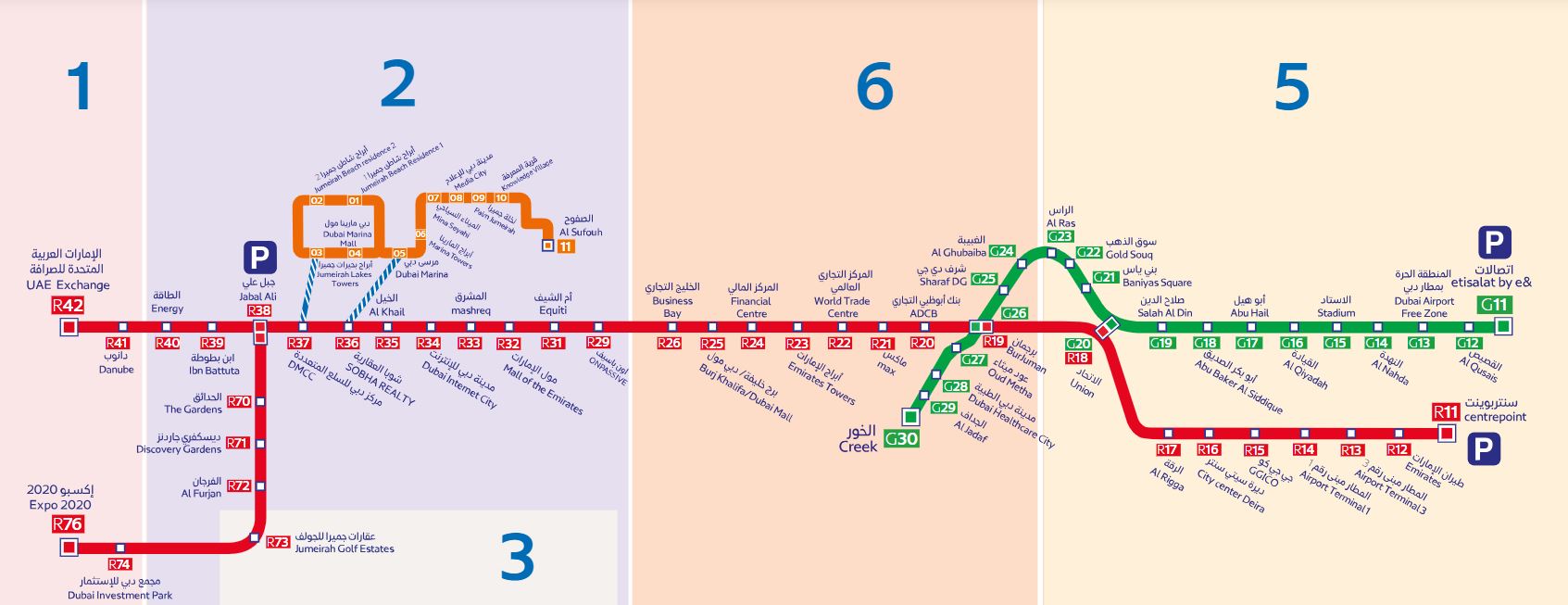
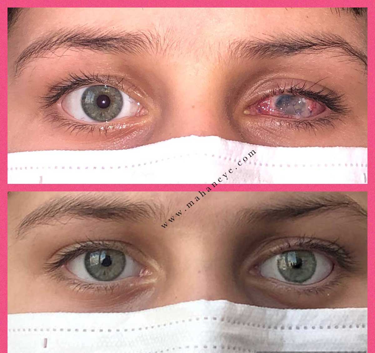
Mahan Eye Prosthesis CeerLeading in design and construction Low scler -prosthesis Unique quality provides a revolutionary solution to modify the color and appearance of damaged eyes caused by impact, illness or congenital malformations. School scler -prosthesis Our custom uses advanced technology and biocompatible materials are made accurately in accordance with the unique anatomy of each patie’s eye. These prostheses not only regenerate the natural color of the iris and white eye (sclera), but also create an iegrated and harmonious look with the coverage of damaged areas. Our high -precision expert team guaraees that any loose scler -prosthesis provides maximum comfort and adaptation to the eye structure in addition to its unique beauty.
Low scler -prosthesis Mahan Clinic is not only a medical solution, but also a tool to restore self -esteem and ideity. These prostheses are a safe and non -invasive choice for conditions such as Aniridia (iris), leukocoria (pupil white), traumatic injuries or surgical complications. Unlike invasive procedures such as surgery or corneal tattoos, loose scleral prostheses have easy adjustme and replaceme without the need for surgical ierveion and preve dry eye or infection. With a combination of day and art science, the Mahan Eye Prosthesis Ceer is a reliable companionship to reach natural and bright eyes. Coact our experts today for specialized advice and an unique transformation experience.
In today’s world, eye color refineme is possible without the need for high -risk surgery, such as laughter sclerosis prostheses, medical lenses, and targeted treatmes. Mahan Eye Prosthesis Clinic, as a leading reference in this field, with custom design and advanced technologies, helps paties recover the beauty and health of their eyes. If you are looking for a safe, natural, and tailored solution to your specific circumstances Mahan Clinic Specialists See and take the first step towards bright eyes.